
Photoacoustic Imaging
Photoacustic imaging (PA) integrates the sensitivity of optical imaging with the resolution of high-frequency ultrasound, to provide insights into tissue microenvironment and hemodynamic changes, addressing the needs of different biological research areas. This hybrid imaging modality is able to detect and quantify endogenous photoacoustic signals of oxy- and deoxy-hemoglobin, as well as photoacoustic contrast agents like gold nanorods and carbon nanotubes that have emerged in recent years as specific and customizable agents for cancer detection and marking of genetic mutations cancer-associated, improving early detection and treatment of neoplasias.
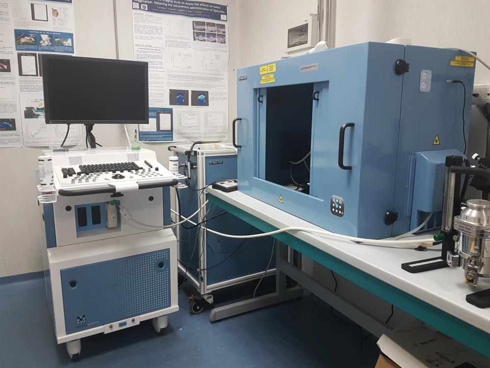 This Photoacoustic Imaging platform allows multispectral imaging with multiple wavelengths (680-970 nm), to detect and quantify signals of oxy- and deoxy-hemoglobin as well as fluorescent dyes with high-resolution, high sensitivity and specificity – even in deep tissues – with real-time and 3D imaging capabilities. Vevo LAZR gives researchers the opportunity to study a wide range of animal models from embryos to adults, with the advantages of inherent co-registration in both 2D and 3D planes of photoacoustic and anatomical images, facilitating biomarker development and longitudinal studies in translational research.
 Available at the centre for Preclinical Imaging in Colleretto Giacosa, at IFC-CNR in Pisa and at IBB-CNR in Naples.

![]()
![]()